Troubleshooting tips for IHC common problems:
1. Non-specifc staining
2. No staining
3. Weak staining
4. Strong Staining
| Causes | Solutions |
| Improper preparation of sections | Improve ways of sampling and preparation |
| Inadequate deparaffinization of the sections | Increase the deparaffinization time |
| Tissue contains endogenous peroxidase | Use 0.3% v/v fresh H2O2 for blocking and increase the blocking incubation time |
| Tissue contains endogenous biotin | Use IHC biotin blocking agent |
| Blocking of protein may be insufficient | Increase the blocking time |
| Charge adsorption | Block with nonimmune animal serum |
| The antibody is not pure | Change suitable antibody |
| Primary antibody concentration may be too high | Try decreasing the antibody concentration |
| The sections have dried out | Avoid sections being dried out in the process of experiments |
| Washes may be insufficient | Increase the times of washes and the washing time |
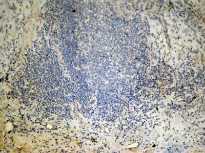
| Non-specifc staining: | Improved: |
 |
 |
Immunohistochemical analysis of paraffin-embedded human lung carcinoma tissue showing cytoplasmic and nuclear staining using NFκB-p65 Phospho-Ser276 Antibody #AB11011.
| Causes | Solutions |
| Improper tissue processing | Try to improve the condition and sampling again |
| No antigen in the tissue | Set a positive control to verify the experiment results |
| The antibody is not active | Don’t use out-of-date antibody kits |
| Incompatible secondary and primary antibodies | Use secondary antibody that was raised against the species in which the primary was raised |
| Incompatible staining system | Change compatible staining system |
| Improper operation and leave out important steps | Follow strict operating procedure and set a positive control |
| No staining: | Improved: |
 |
 |
Immunohistochemical analysis of paraffin-embedded human breast carcinoma tissue using Histone H3 Di-Methyl-Lys27 Antibody #AB11583.
| Causes | Solutions |
| Improper tissue fixation or too high temperature when fixing | Use appropriate fixation way or fixation time |
| Too high baking slides temperature and too long baking time | Choose appropriate temperature and time for baking slides |
| The antigen may be damaged | Let fresh tissues be fixed in time and for not to exceed 24 hours |
| Over blocking of protein | Reduce the blocking time |
| The antibody has drained away | Ensure that the sections are placed in a horizontal position when incubating |
| The antibody concentration may be too low or incubation time may be too short | Increase the antibody concentration and incubation time |
| The room temperature may be too low. Lower than 15℃. | Incubator at 37℃ or increase incubation time |
| No draining off buffer solution when adding the reagent results in the reagent being diluted. | Do drain off buffer solution but avoid sections being dried out. |
| Excessive washing | Wash moderately |
| Always verify the expiration date of the reagent prior to use | Change reagents timely |
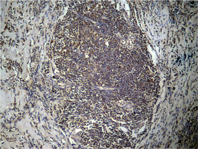
| Weak staining: | Improved: |
 |
 |
Immunohistochemical analysis of paraffin-embedded human breast carcinoma tissue using mTOR Phospho-Ser2448 Antibody#AB11221.
| Causes | Solutions |
| The primary antibody concentration may be too high or incubation time may be too long | Reduce the primary antibody concentration or incubation time. |
| The incubation temperature may be too high | Incubate at 4℃ or at room temperature |
| The incubation time of HRP conjugated secondary antibody may be too long | Reduce the incubation time |
| Inadequate washing | Increase the times of washing |
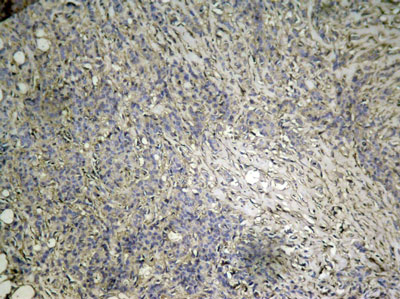
| Strong Staining: | Improved: |
.jpg) |
.jpg) |
Immunohistochemical analysis of paraffin-embedded human breast carcinoma tissue using FKHR Phospho-Ser256 Antibody#AB11115.
© 2017, AbSci All Rights Reserved. E-mail: info@abscitech.com
南京川博生物技术有限公司版权所有
苏B1-20150380 苏ICP备15009006号-1